In a new paper recently published in the journal PNAS, Gayani Senevirathne, a graduate student in the Shubin Lab, has described fascinating new work on the ontogeny of the anuran urostyle and the developmental context of evolutionary novelty (click here to read the paper).
Significance of this work:
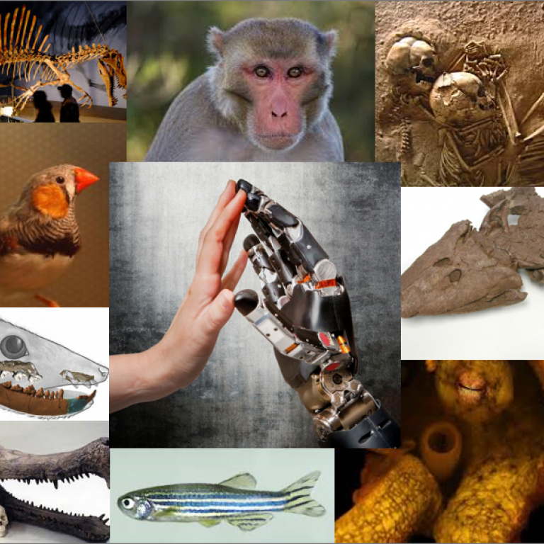
Fusion of caudal vertebrae has evolved multiple times independently: the pygostyle of birds, coccyx in apes and humans, ural plate of fish, and the urostyle of frogs. The anuran urostyle, however, is structurally and developmentally distinct because of the contribution of an ossifying hypochord. To date, the developmental mechanisms behind an ossifying hypochord have remained obscure. Here, we provide a detailed analysis of the development of this evolutionary innovative structure and of how neuromusculature, cell death, and proliferation paved their way to facilitate its formation. Finally, we propose that the ossifying hypochord plays a role in tail loss in anurans and reorganizing the dorsal aorta and thus is pivotal in the evolution of the anuran bauplan.
To understand the evolution of the urostyle, Senevirathne focused on the hypochor, a rod-like column of cells that develops below the notochord in amniote vertebrates. Early development of the hypochord and its signaling properties and responses have been described in early development of zebrafish.The salamander hypochord is apparently transient, because Löfberg and Collazo report its development and its position and potential influence on the dorsal aorta but report disappearance after 8 days, owing to extensive apoptosis. Wake and Wake found no evidence for a hypochord in the caecilian Dermophis mexicanus in their examination of early vertebrogenesis and the notochord (perhaps correlated with the absence of girdles, limbs, and tail). Senevirathne specifically assessed the origin of the ossifying hypochord in order to assess its origin, its association with the notochord (physically and in signaling), with the terminal vertebrae (the coccygeal component of the urostyle), with the dorsal aorta, and with the process of metamorphosis. They parsed the questions about the hypochord in terms of problem and of technique, employing both “traditional” and new genetic tools. Clearing and staining (alizarin red S for bone and Alcian blue for cartilage) a series of whole tadpoles was used to develop a staged series of embryos and tadpoles for examination of the development of the cartilage and bone of the vertebral column, the urostyle, and limbs. Histological preparations revealed the cell structure of the developing structures. Maintaining stage-54 tadpoles in a solution containing thyroxin and a control series in a solution without thyroxin, then raising them for 2 months, finally clearing and staining them, was used to assess the effect of thyroxin on urostyle development. A series of tadpoles at stage 54 were stained with phosphomolybdic acid and the specimens were scanned by computed tomography to follow development of the urostyle. Scans were analyzed and segmented. Six tadpoles of each of four stages were immunohistochemically stained to examine cell death, neurons, and muscle remodeling. Antibodies used included Caspase-3 to observe apoptosis, acetylated tubulin for neurons, and Laminen for muscle fibers. Whole-mount in situ hybridization and whole-mount immunohistochemistry were done with a diversity of enzymes and antibodies. The abundant illustrations in the publication reflect the copious body of specimens which were examined and data taken and analyzed.
(Commentary excerpted from PNAS)